
Key Takeaways
- The axial skeleton forms the central framework, supporting and protecting vital organs including the brain, heart, and lungs.
- The appendicular skeleton consists of the limbs and girdles, facilitating mobility and interaction with the environment.
- The axial skeleton is primarily responsible for structural stability, while the appendicular skeleton enables movement and manipulation.
- Both skeletal divisions work in concert to maintain posture and enable complex physical activities.
- Distinct anatomical features and functions differentiate the axial and appendicular skeletons, reflecting their unique roles in human biomechanics.
What is Axial Skeleton?

The axial skeleton is the core structural system of the human body, encompassing bones along the central axis. It plays a critical role in protecting essential organs and supporting the weight of the head and trunk.
Components and Structure
The axial skeleton includes the skull, vertebral column, ribs, and sternum. These bones form a protective case around the brain, spinal cord, and thoracic organs, ensuring their safety from external trauma.
The vertebral column itself comprises 33 vertebrae, segmented into cervical, thoracic, lumbar, sacral, and coccygeal regions. This segmentation allows for both rigidity and flexibility necessary for upright posture and movement.
The rib cage, formed by 12 pairs of ribs and the sternum, encloses the lungs and heart, providing mechanical protection and supporting respiratory function. Additionally, the rib cage allows expansion and contraction during breathing, illustrating its dynamic role.
Role in Posture and Stability
The axial skeleton acts as the central support pillar for the body, maintaining posture by bearing the weight of the head, neck, and trunk. Without this support, balance and upright positioning would be compromised.
The vertebral column’s curvature helps distribute mechanical stress during movement, reducing injury risks. This natural alignment also absorbs shock when walking or running, protecting internal structures.
Muscle attachment sites along the axial skeleton provide leverage for maintaining stability, particularly around the spine and thoracic cage. These attachments facilitate fine motor control essential for everyday activities such as sitting and standing.
Protection of Vital Organs
The skull encases the brain within a rigid bony vault, shielding it from impact and injury. Its complex structure includes various bones joined by sutures that accommodate growth and minor flexibility.
The vertebral column protects the spinal cord, a critical conduit for nerve signals between the brain and the rest of the body. Damage to this area can result in severe neurological impairments, highlighting its protective importance.
The thoracic cage safeguards the heart and lungs, organs essential for circulation and respiration. Its semi-rigid design balances protection with the need for expansion during respiratory cycles.
Development and Growth Patterns
During human development, the axial skeleton forms early and establishes the body’s primary framework. Ossification centers in the vertebrae and skull appear prenatally, setting the stage for postnatal growth.
The vertebral column undergoes changes in curvature during infancy and childhood, reflecting the acquisition of motor skills such as sitting and walking. These curves improve biomechanical efficiency in adult posture.
Bone remodeling in the axial skeleton continues throughout life, adapting to mechanical stress and repair needs. This dynamic process ensures the skeleton remains functional despite daily wear and tear.
What is Appendicular Skeleton?
The appendicular skeleton comprises the bones of the limbs and their corresponding girdles, facilitating movement and interaction with the environment. It plays a vital role in locomotion and manipulation of objects.
Composition and Regional Divisions
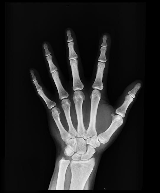
The appendicular skeleton includes the pectoral girdle, upper limbs, pelvic girdle, and lower limbs. Each region has specialized bones adapted for specific functions such as grasping, lifting, or walking.
The pectoral girdle consists of the clavicles and scapulae, connecting the upper limbs to the axial skeleton and allowing a wide range of shoulder motions. Its design prioritizes flexibility and mobility over rigid protection.
The pelvic girdle forms a strong, stable connection between the lower limbs and the axial skeleton, supporting body weight during standing and locomotion. Its robust structure reflects the demands of bearing and transferring load.
Function in Mobility and Interaction
The appendicular skeleton provides the mechanical basis for voluntary movement, enabling activities like walking, running, and grasping. Joints within this skeleton exhibit a wide range of motions, from hinge-like bending to rotational twisting.
Upper limb bones, such as the humerus, radius, and ulna, work together to allow precise hand positioning and tool use. This dexterity is critical for tasks ranging from writing to complex manual labor.
Lower limb bones, including the femur, tibia, and fibula, are adapted for weight-bearing and propulsion. Their strength and alignment are essential for efficient gait and balance, particularly on uneven terrain.
Adaptations for Environment and Activity
Variations in appendicular skeleton morphology reflect environmental and lifestyle influences. For example, populations engaged in heavy physical labor often exhibit more robust limb bones compared to those with predominantly sedentary lifestyles.
Sports and physical training can induce changes such as increased bone density and muscle attachment size within the appendicular skeleton. These adaptations improve performance and reduce injury risks.
In evolutionary terms, the appendicular skeleton has undergone significant modification to facilitate bipedalism and tool use, distinguishing humans from other primates. This evolutionary trajectory underscores the skeleton’s role in survival and cultural development.
Growth and Development Dynamics
Long bones within the appendicular skeleton grow through endochondral ossification at growth plates located near the ends of these bones. This process continues until skeletal maturity, typically in late adolescence.
Growth patterns differ between upper and lower limbs, often reflecting functional priorities such as hand dexterity or walking efficiency. These differences can influence body proportions and biomechanics.
Throughout life, the appendicular skeleton undergoes remodeling in response to mechanical loading and injury, maintaining structural integrity and function. This adaptability is crucial for maintaining mobility in aging populations.
Comparison Table
The following table highlights key distinctions and functional characteristics that differentiate the axial and appendicular skeletons.
| Parameter of Comparison | Axial Skeleton | Appendicular Skeleton |
|---|---|---|
| Primary Function | Protects vital organs and supports body’s central axis | Facilitates movement and environmental interaction |
| Bone Count | Approximately 80 bones | Approximately 126 bones |
| Major Bone Groups | Skull, vertebral column, ribs, sternum | Limbs and girdles (pectoral and pelvic) |
| Joint Types | Primarily immovable or slightly movable joints | Mostly freely movable synovial joints |
| Role in Locomotion | Indirect, by providing support and structure | Direct, enabling walking, running, and manipulation |
| Protection Focus | Encases brain, spinal cord, heart, and lungs | Minimal protection; mainly structural and functional |
| Flexibility | Limited flexibility with structural rigidity | High flexibility to accommodate wide range of motions |
| Muscle Attachment | Supports posture-related muscles | Supports muscles responsible for movement and strength |
| Load Bearing | Supports axial load and distributes weight | Supports dynamic load during movement and balance |
| Development Pattern | Forms early, establishing central framework | Develops later, adapting to functional demands |